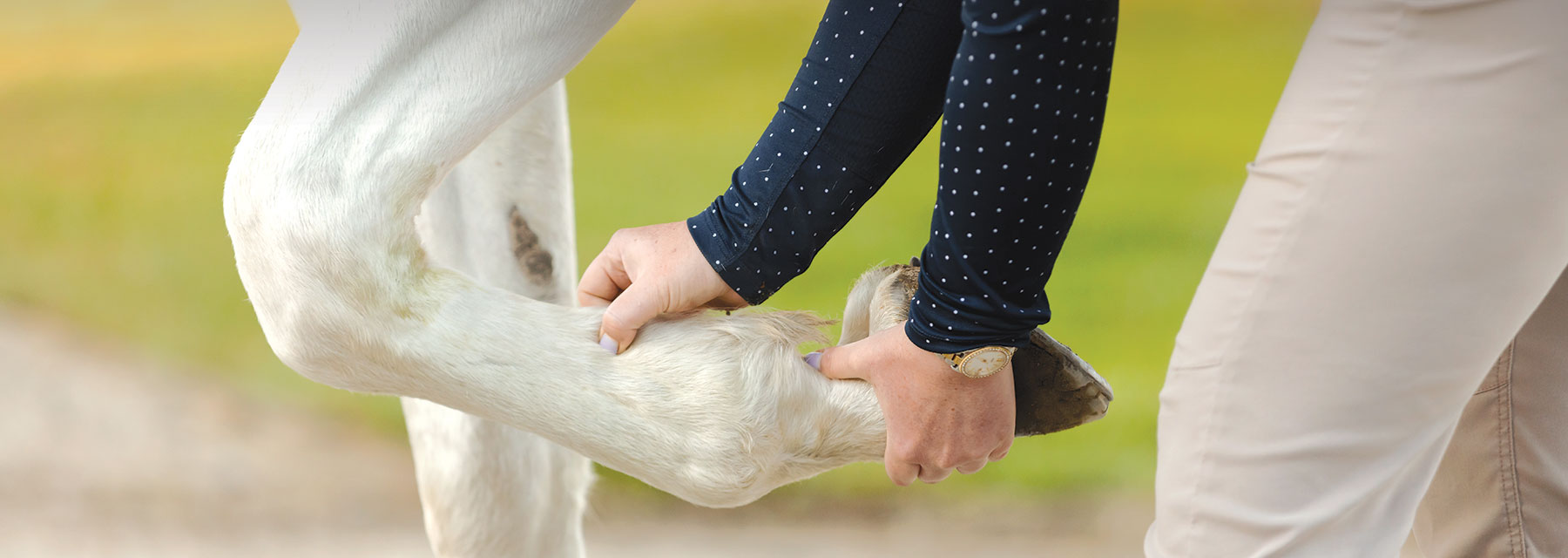
A Step-by-Step Guide Through an Equine Lameness Exam and Treatment Plan Options
Lameness is an alteration of the horse’s gait. While not actually a disease, it is a clinical sign resulting from restrained movement and pain in muscles, nerves, soft tissue, bones or joints. The range of causes include strain or injury, laminitis, infection, metabolic or genetic issues. Less common, neuromuscular disorders that affect a horse’s muscles and nerves can also produce lameness, although usually with a non-painful presentation. Lameness severity runs the gamut from a limp to a limb that cannot bear weight, with subtleties, such as a change in performance or even the animal’s general attitude. Typically thought of as a leg problem, lameness issues can also stem from other parts of the body and include the neck, back or hooves.
Lameness examinations are performed by veterinarians to diagnose the condition and locate the source of the problem. They are also often part of a pre-purchase examination or pre-lease situation to ensure a horse is sound and suitable for its intended job. The exam typically includes a medical history, physical exam, visual and in motion observation, flexions — or the bending of a limb, diagnostic testing and a treatment plan. This exam can be time consuming particularly when the cause is not obvious, requiring a process of elimination to identify the causative factor.
“We call them lameness exams, but really, it’s more of a performance evaluation or even a movement evaluation. ... As veterinarians, we have to first identify the specific problem, then match an appropriate treatment plan to return the patient back to full athletic capability whatever that may entail — whether it’s a Grand Prix jumper or my kid’s pony.”
— Dr. Clayton Smith, Owner of Smith Equine Performance & Rehabilitation in Cypress, Texas
Parts of a Lameness Exam
Medical History
A veterinarian needs to know the general demographics of the horse: breed, sex, age and past or present health issues. If the horse is in work, the discipline and training schedule should be discussed. Is the rider or trainer feeling anything unusual under tack? Often riders feel subtle changes that could be important clues to the diagnosis. The shoeing cycle is important to know — especially shoeing changes. The vet should know when the lameness was noticed and if it has responded positively to treatments, such as rest, exercise or anti-inflammatory or analgesic medications. The veterinarian must be alerted if any analgesic has been given before the exam. Any recent bloodwork or biochemical analyses should be shared if an infectious, metabolic or myopathy issue is suspected.
Physical Exam
Ask your veterinarian ahead of the visit how they will first evaluate the horse. Most prefer to evaluate the horse “cold,” without having been exercised, but in some circumstances, examining a warmed-up animal is helpful. At times, some begin by observing the horse free in its stall. A veterinarian wants to see how the horse is bearing weight on each limb. The horse should be restrained and standing evenly on a level surface. General conformation, symmetry and balance are evaluated, looking for muscle loss or an asymmetrical stance. Manual palpation is used to check muscles, soft tissue, bones and joints for any indication of pain, heat, swelling, thickening or other abnormalities.
Hooves will be thoroughly examined. Hoof testers that apply pressure and compress the sole and frog of the hoof are used to check for sensitivity and pain. Your vet will look for wear patterns on the shoes or feet, and abnormalities, such as mismatched hoof angles, contracted or sheared heels will be assessed. Irregular or disproportionate hoof sizes can also cause lameness or, conversely, can be evidence of uneven limb loading. Horseshoes are usually only removed when the cause is localized to the foot.
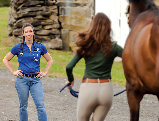
In most circumstances, observing a horse in motion is an essential part of a lameness exam. A horse is typically examined on a level, nonslip surface at a walk and trot in both directions on a straight line and in a circle. The veterinarian is looking for any deviations in movement — a shortened stride, head bobbing, shifting weight, stiffness or any inability to land each foot evenly.
Visual/In-Motion Exam
In most circumstances, observing a horse in motion is an essential part of a lameness exam. A horse is typically examined on a level, nonslip surface at a walk and trot in both directions on a straight line and in a circle. The veterinarian is looking for any deviations in movement — a shortened stride, head bobbing, shifting weight, stiffness or any inability to land each foot evenly. Often, lameness becomes more obvious when the horse works in a circle, which can be done in hand, on a lunge line, in a round pen or under saddle. Ideally, the same handler is used throughout the exam. When jogged, a horse should be controlled, so the vet can better see the animal moving in a consistent, repeatable pace. Some gait analysis systems are available to quantify lameness and help with detection.
It may be beneficial to watch the horse being lunged in a circle around the handler or ridden under tack. Under saddle evaluation can be helpful particularly for subtle lamenesses or those only felt while riding or seen in weight-bearing instances. The horse should be ridden in its usual bridle and tack and on a softer surface, such as in an arena. This can make it easier to identify a multiple-limb lameness as opposed to the more obvious single-limb lameness. Clinical signs of discomfort can include the horse’s unwillingness to perform certain movements or activities, tail wringing, head tilting, etc. It should be noted that some clinical signs are, in fact, training or behavior problems not associated with lameness. Ruling out lameness or pain in the horse should always be done first for any behavioral issue. Severity of lameness will dictate how extensive the in-motion part of the examination is.
The most obvious sign of unilateral forelimb lameness is the head bob when the head and neck lift when the lame limb bears weight as it meets the ground. The head and neck fall with a sound limb. For hindlimb lameness, the pelvic or sacral rise can often be seen in a similar fashion. The pelvis will rise when the lame limb bears weight and touches the ground, and, conversely, will fall when the sound limb strikes. The reason is that the rising of the weight reduces the concussive force on the lame leg.
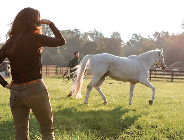
Often, lameness becomes more obvious when the horse works in a circle, which can be done in hand, on a lunge line, in a round pen or under saddle.
Often, lameness becomes more obvious when the horse works in a circle, which can be done in hand, on a lunge line, in a round pen or under saddle.
The AAEP Lameness Scale
- Lameness is not perceptible under any circumstances.
- Lameness is difficult to observe and is not consistently apparent, regardless of circumstances (e.g., under saddle, circling, inclines, hard surface, etc.).
- Lameness is difficult to observe at a walk or when trotting in a straight line, but consistently apparent in certain circumstances (e.g., under saddle, circling, inclines, hard surface, etc.).
- Lameness is consistently observable at a trot under all circumstances.
- Lameness is obvious at a walk.
- Lameness produces minimal weight-bearing in motion and/or at rest, or a complete inability to move
Flexions
A flexion test stresses different parts of the leg to isolate the problem area. The veterinarian watches the horse trot in straight line back and forth as a baseline. For the flexions, the vet holds up the leg in a tight stretch for a consistent period and wants to watch the horse trot immediately after the limb is released, noting changes in movement. The flexion pressure must be consistent and firm but not excessive, which can cause a false positive. Standard flexion tests are carried out in a straight line on a hard surface with joints flexed one at a time. Flexions are done on sound and lame limbs for comparison. There are essentially two parts to this test. In the passive portion, while the limb is held in flexion, range of motion and any induced discomfort is assessed. In the dynamic phase, increases in lameness indicate limb stress. Most commonly, positive flexion tests reflect joint involvement while other issues can also be exacerbated through bending of the limb.
The American Association of Equine Practitioners (AAEP) uses a five-point scale of motion and flexion lameness with one indicating the condition is difficult to observe and five as a complete inability to bear weight. (Zero indicates no lameness is observed. The AAEP lameness scale is subjective.)
Diagnostic Tests
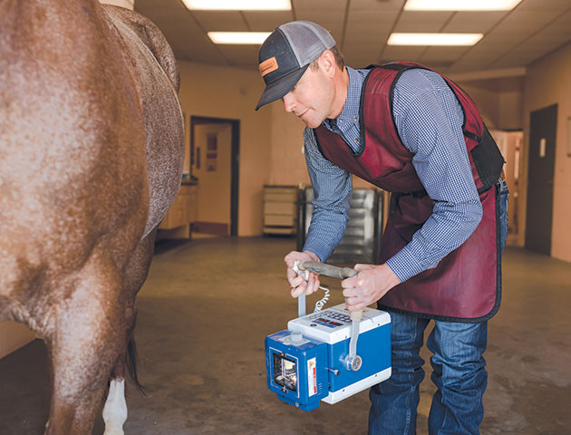
Diagnostic testing is used to pinpoint the specific cause of pain. These tests may include digital palpation, nerve blocks, joint blocks, radiographs, ultrasound, MRI, nuclear scintigraphy (bone scan) and lab tests.
After the physical and visual exams and flexions, a veterinarian will have determined which limb (or combination of limbs) is causing the lameness. Diagnostic testing is used to pinpoint the specific cause of pain. These tests may include digital palpation, nerve blocks, joint blocks, radiographs, ultrasound, MRI, nuclear scintigraphy (bone scan) and lab tests. For nerve and/or joint blocks, an anesthetic injection numbs the nerves around specific areas of the leg to determine the area of pain whenever the lameness can be localized to a specific limb but not a specific site on the leg. This helps zero-in on the source of the pain. Once the area is numb, or blocked, the horse is jogged to compare if he is sounder without being able to feel the problem area. If the horse is sounder with the block, then that is likely the area causing pain.
Radiographs or X-rays (electromagnetic radiation) is non-invasive imaging used to identify damage or changes to boney tissues. Several images will be taken of different angles of a joint. The AAEP recommends four images for assessing a single joint. Ultrasound images are very helpful for suspected soft tissue damage in tendons, ligaments or structures within the joint and can show the surfaces of bone well. This non-invasive technology uses high-frequency sound waves to create pictures of what it looks like inside the body. Magnetic resonance imaging, or MRI, is sometimes required for difficult cases or difficult body regions to image with X-ray or ultrasound, such as the hoof or proximal suspensory region. This advanced diagnostic tool uses radio waves and a powerful magnet to generate detailed images. As an MRI is not readily available in most individual veterinary clinics, the horse may be referred to a hospital or other facility that has an MRI to have the test performed.
Bloodwork, synovial (joint) fluid or tissue samples may be sent to labs to test for markers of infection or inflammation.
While basic neurologic assessment is noted during the initial exam, a more thorough neurologic review may become part of a lameness exam in cases where pain or locomotor causes of lameness cannot be located. If a neuromuscular disorder is suspected, the vet may want to observe the horse doing specific movements, such as backing, negotiating a curb, turning in tight circles and walking up and down a slope to evaluate spatial awareness, weakness or gait abnormality. Cranial nerves and upper and lower motor neuron function will be evaluated if a neurologic deficit is considered.
Treatment Plan
Once the lameness source is identified, a personalized treatment plan can be developed for the patient. The individualized treatment plan can include the horse’s history, specific injury and performance goals. A major focus of lameness therapies is often to relieve or mitigate inflammation. Inflammation is a natural and expected part of physiology and, within normal limits, is considered healthy as it stimulates repair and even provides disease protection. However, an overactive inflammatory response — especially over an extended period — lead to tissue destruction.
While many factors contribute to lameness, issues affecting the joint or surrounding tissues are the most common reasons a lameness exam is needed. Equine joints can be affected by acute and chronic inflammation. The latter can be particularly problematic with the potential to overwhelm the natural systems that control it. Repetitive wear and tear, as is seen with chronic types of inflammation, leads to the cartilage breakdown, most commonly occurring in the weight-bearing and high-motion joints of the legs — the knees, hocks, stifles, fetlocks and the coffin joint in the hoof. The joints within the spine also can be affected.
Anti-inflammatory support will be a part of, if not the entire, focus of treatment. Cold therapy, like icing, is a simple and effective first step to reduce acute inflammation. Prescription medications, such as NSAIDs, non-steroidal anti-inflammatory drugs like phenylbutazone (bute) or flunixin meglumine (Banamine®), focus on anti-inflammatory support as well as pain control to support comfort and healing. They are typically prescribed shortterm because of the gastrointestinal and kidney issues found with long-term use.
Dependent on the source of the lameness, intra-articular joint injections using corticosteroids, hyaluronic acid or a combination can help re-establish lubricating properties and decrease inflammation within the joint capsule. Horses receiving joint injections often have fast-acting and acute support as the synthetic lubricating fluids are injected directly into the joint capsule. The often-used intramuscular Adequan™ and intravenous Legend™ are considered chondroprotectants, working either directly on the cartilage or on the synovial fluid within the joint respectively, with the primary objective of preventing or delaying cartilage issues. Therapies that focus on using the body’s own system to stimulate regeneration and healing may be advocated. Examples include interleukin receptor antagonist protein (IRAP), platelet rich plasma (PRP), Pro- Stride®, stem cell therapy and shockwave. Shoeing changes may be needed to support certain inflamed areas of the foot or soft tissues of the leg and facilitate healing. Veterinarians often work closely with farriers to find the best shoeing option for each case that supports the natural anatomy and offers comfort for the specific lameness.
Depending on the injury, rehabilitation plans may be needed to slowly rebuild muscle and put gradual, controlled stress on the source of the lameness. Working within individual management parameters, a veterinarian will create a timeline for returning the horse to work, if possible. If your horse is recovering from an injury and rehabilitation seems overwhelming, dedicated equine rehab facilities are an option. Many offer a dry or underwater treadmill, horse walker, cold saltwater spa, swimming lane, vibration plate, magnetic blankets and more. Adjunct therapies — acupuncture, chiropractic or massage — may be recommended. The animal’s general diet should support reducing inflammation by limiting or eliminating cereal grains and sugar. Supplementation with omega-3 fatty acids, antioxidants and joint-specific nutrients support wellness and a healthy recovery.
One of the very best things to do is simply resting your lame horse. Inflamed tissues require time to calm down and repair without the strain of performance or ongoing wear and tear. Time off from formal exercise is almost always recommended for lameness and is often the cornerstone of a treatment plan. A veterinary re-check will likely be recommended to monitor progress and adjust the treatment as needed.
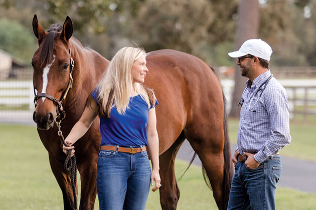
“The challenges that go with being an equine lameness specialist is what gets me out of bed in the morning. The subtle lamenesses are extremely fascinating to me. ... Using the different modalities available to help diagnose and treat equine athletes and help me perfect my craft.”
— Dr. Clayton Smith, Smith Equine Performance & Rehabilitation
What is a Soundness Exam?
Many veterinarians recommend performing periodic soundness examinations, which is essentially the same as a lameness examination but when no lameness is noted. The exam can often uncover subtle issues that are sometimes more easily addressed before they turn into a full lameness issue.
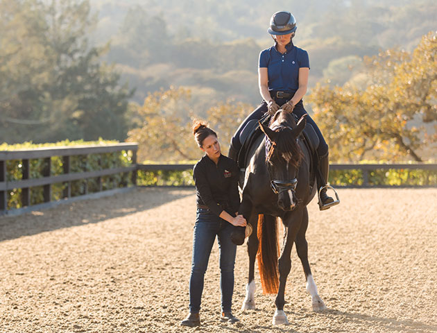
Many veterinarians recommend performing periodic soundness examinations, essentially the same as a lameness examination but when no lameness is noted. The exam can often uncover subtle issues that are sometimes more easily addressed before they turn into a full lameness issue.
Supplementation to Support Soundness
Daily supplements allow nutrients to be consistently available to the body and actively working to support the many aspects of the intricate equine anatomy. Once starting a supplement, full cellular saturation should be accomplished after 30 days, although anecdotal benefits may occur prior to then.
To address lameness concerns, a comprehensive joint supplement provides the best support as different ingredients may offer support to different parts of the body as needed. Specific nutrients support healthy joints, soft tissue and overall wellness. Ingredients in a supplement serve one or more of these purposes: to support normal inflammation; to help promote cartilage synthesis and other joint components; or to promote normal levels of certain enzymes that degrade joint tissue. For instance, glucosamine, is known to both support normal levels of inflammation and is a natural building block for cartilage cells. These supplements include:
- Glycosaminoglycans (GAGs) are long, unbranched polysaccharides that are highly polar and attract water, making them critically useful in the body as a lubricant and shock absorber. GAGs form an important component of connective tissues and are key to normal, healthy joint function. Glucosamine is an amino sugar that can be a building block for GAGs and cartilage. It may promote the production of new GAGs and help maintain normal healthy cartilage.
- Hyaluronic acid (HA) is a GAG and a major component in the synovial fluid lubricant in body joints and soft tissues. HA is present within joints to reduce friction as well as absorb shock between bones during movement. In doing so, it helps maintain healthy joint function. Although it is commonly administered intra-articularly via joint injections, orally administered HA has been shown in several studies to be bioavailable and effective in supporting joint health. HA is often used to support existing joint conditions or for horses subjected to activities that can lead to joint concerns.
- Methylsulfonylmethane (MSM) is a sulfur-containing compound and product of DMSO (Dimethyl Sulfoxide) that is often advocated for joint support. MSM plays a significant role in joint health by providing a bioavailable sulfur source that is a key component in most GAGs, cartilage, tendons and ligaments. Animal studies have shown MSM to help maintain healthy skin, coat and hoof quality, and it is necessary to support the formation-reinforcing bonds between collagen strands. MSM has been shown to support healthy levels of both inflammation and oxidative stress in the horse.
- Cetyl myristoleate is a naturally derived ester, or organic compound, of an omega-5 fatty acid that helps maintain joint health by working at the precise location of joint inflammation. It is effective in supporting joint discomfort and promoting mobility and joint functionality.
- Omega-3 fatty acids are essential fatty acids, indicating that they are not produced by the body and therefore must be consumed in the diet. Omega-3s are important for cellular health and can help support normal, healthy levels of inflammation in the body, as well as help maintain joint health and multiple other aspects of wellness. Most important to the joint health is that dietary omega-3 fats have the ability to inhibit aggrecanase, an enzyme involved in degrading proteoglycan content in articular cartilage.
- Free radicals are chemicals produced in a horse’s body either as a result of normal metabolism, or in response to exercise, oxygen, inhalation of dust and air pollutants, ingestion of rancid feeds or exposure to ultraviolet light. Free radicals can damage cell membranes and produce lesions in joints and several other tissues. Antioxidants in the horse help neutralize free radicals under normal conditions. Supplementation is warranted to deal with elevated levels of free radicals that can lead to oxidative stress related to exercising horses, horses with health conditions or other variations of bodily stressors or for animals on a high grain diet. Oxidative stress is another culprit for joint aggravation. Vitamin C (ascorbic acid) is a water-soluble vitamin with strong antioxidant actions, protecting against free radical attack. Vitamin C is required for the development of cartilage and the formation of collagen, making it much needed for healthy joints and connective tissue. Vitamin E is a potent antioxidant and, in concert with vitamin C, reduces the damage of free radicals and is often supplemented for this purpose.
“The more we learn about lameness diagnostics and treating horses, especially athletes, the more we realize it’s a whole-horse approach. This includes genetics, fitness, training, veterinary care, farriery and diet. Recovery is crucial for equine athletes, and if they don’t have the nutritional support needed to heal from the inside for growth and repair, you’re going to run into problems.”
— Dr. Clayton Smith, Smith Equine Performance & Rehabilitation
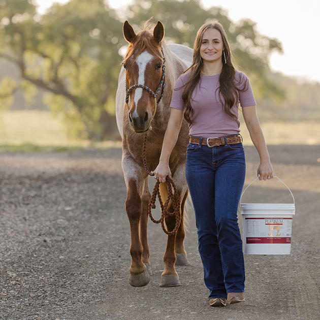
Platinum's most potent joint combination is Platinum Performance® Equine with Platinum Joint Care + HA.
Platinum's most potent joint combination is Platinum Performance® Equine with Platinum Joint Care + HA.
Which Platinum Formulas Should I Use?
The combination of Platinum Performance® Equine and Platinum Joint Care + HA provides joint-focused ingredients like glucosamine sulfate, MSM, cetyl myristoleate and hyaluronic acid with a whole-horse wellness formula that includes omega-3 fatty acids and antioxidants supporting every part of the horse from head to hoof.
It is Platinum’s most potent combination for joint support and is popular with performance horses, senior horses and horses recovering from lameness or injury because it addresses joint health from all angles.
Does My Horse Need a Supplement if the Vet Injected His Joints?
Oral joint supplements work differently than joint injections as supporting ingredients are ingested on a daily basis, pulled into the blood system and available for use by the body when needed. It is a different and generally considered safe option that provides support to the horse. Used independently or in combination with pharmaceutical joint therapies, oral supplements can be a less invasive, longterm way to provide joint support. Most joint supplement ingredients offer some support on the ubiquitous inflammation that most horses deal with on some level and at some point. Specific joint-targeted nutraceutical ingredients that exhibit health benefits and therapeutic value can be used for all ages and life stages of horses and are almost synonymous with athletic support supplements for competition horses as well as senior horse supplements.
